
Fig. 3.
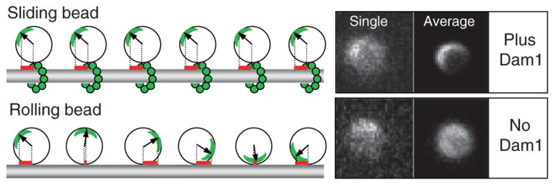
Dam1-coated beads can move via two different mechanisms depending on the availability of soluble Dam1. Left panel illustrates that when bead slides, a projection on the MT axis of the vector from bead’s center to the fluorescent mark (red bars) does not change, while in the rolling bead this measure changes significantly. Right panels show images of two 1-μm Alexa488-Dam1-coated beads that tracked the shortening MT ends in our assay. Both beads had a fluorescent mark, as seen on their “single” images (one frame from the corresponding sequences). The bead on top moved without rolling (soluble Dam1 was present), so when all images in this sequence were centered and averaged, a fluorescent crescent became highly visible. Lower “average” image is for the bead that tracked shortening MT end in a buffer with no