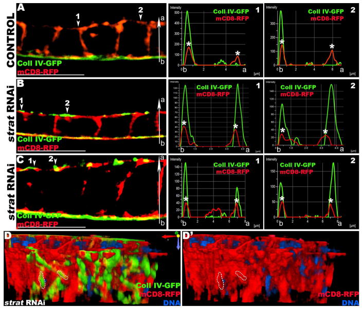
Figure 2. Stratum depleted FCs secrete BM proteins apically.
(A–C) 3D-SIM reconstructions and micrographs of Lg-sections through a control (A) or strat RNAi (B, C) FE expressing Coll IV-GFP (green) and the plasma membrane marker mCD8-RFP (red) exclusively in the FCs. The distributions of green (Coll IV-GFP) and red (mCD8-RFP) pixels along the basal-apical axis (b-a) at different positions of the epithelium (arrowheads) are plotted in histograms to the right of the corresponding micrograph. The x-axis represents the distance along the b-a axis (μm); the y-axis represents arbitrary pixel intensity. The basal and apical plasma membranes are marked with asterisks (*). (A) In control FE, Coll IV-GFP is only secreted at the basal side of FCs and its tightly associated with the basal plasma membrane. Histograms show the distribution of pixels along the b-a axis (red and green pixel peaks overlap). (B–C) In strat RNAi cuboidal (B) or columnar (C) FCs, Coll IV accumulates on both apical and basal sides. At the apical side, Coll IV and the plasma membrane are closely associated, although distinct layers (arrowheads). Histograms, showing the distribution of pixels along the b-a axis, reveal that Coll IV is tightly associated with both basal and apical plasma membranes (red and green peaks overlap on both sides) and accumulates apically outside of cells (green peaks detected apically to red peaks). (D) 3D reconstruction of a strat RNAi FE (same as C). View facing the apical side of the FE. Orientation reference is shown. Coll IV covers the apical FE plasma membrane, indicating that Coll IV is secreted apically outside of the cell (see dotted lines). Bars, 10 μm.

