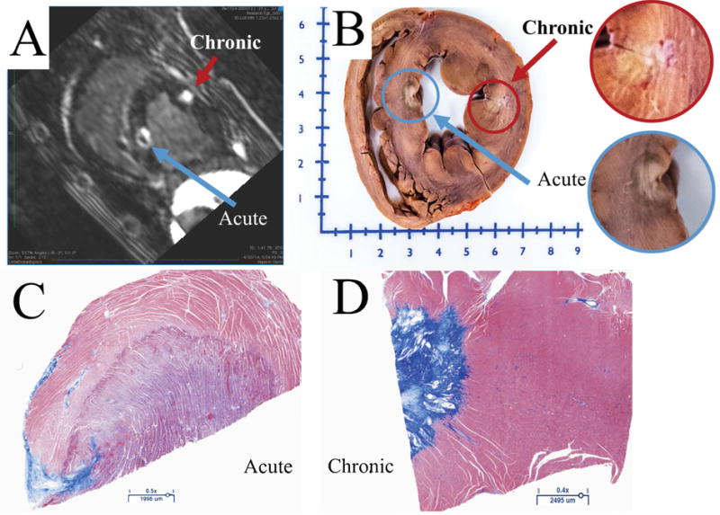
Figure 2.

A) Short axis view from LGE-MRI scan showing acute and chronic RF lesions. The chronic lesion is characterized with a single enhanced area, while acute lesion consists of three: MVO, enhanced and edema areas. B) Gross pathology showing acute and chronic lesions. C) The histology of an acute lesion. D) The histology of a chronic lesion with Masson’s Trichrome staining.
