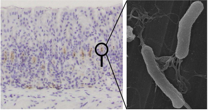
Figure 1.
Chronically Helicobacter pylori infected mouse stomach and electrion microscopic image of H. pylori. (Left) Light micrograph of H. pylori detected by immunohistochemistry (Thermo Fisher Scientific, Fremont, CA) (brown dots) in the stomach of CD1 mouse, 6 months after H. pylori inoculation. Note that inflammation and tissue damage were not detected despite the presence of H. pylori. (Right) Electron micrograph of H. pylori with the spiral shape and multiple flagella. Image was taken by an ultra-high resolution scanning electron microscope S-900 (Hitachi, Ibaraki, Japan) at Kindai University Faculty of Medicine (Osaka, Japan).

