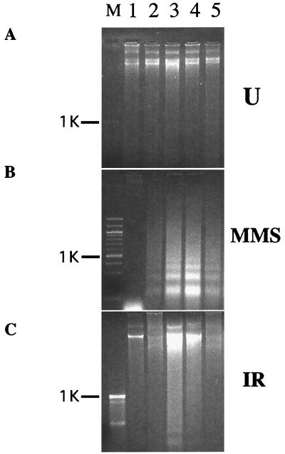
Figure 4.
DNA fragmentation apoptosis assay following DNA damage in cells expressing GADD34 and Lyn. Agarose gel electropherograms of DNA from HEK293 cells transfected with 5 μg each of pcDNA3.1 and pFLAG-CMV2 (lanes 1 and 2), pFLAG-CMV2-GADD34 + pcDNA3.1 (lane 3), pFLAG-CMV2 + pcDNA3-Lyn (lane 4), pFLAG-CMV2-GADD34 + pcDNA3-Lyn (lane 5), and either left untreated (A) or treated with MMS (B) or IR (C). FLAG-GADD34 and Lyn expression levels were equal in double and single transfectants, as judged by Western blots (data not shown). DNA was extracted 12 h following treatment because at this time point, DNA laddering is most pronounced (data not shown). M, molecular size marker lane; position of 1-kb band is indicated. Two other repeats of the same experiment produced similar results (data not shown).

