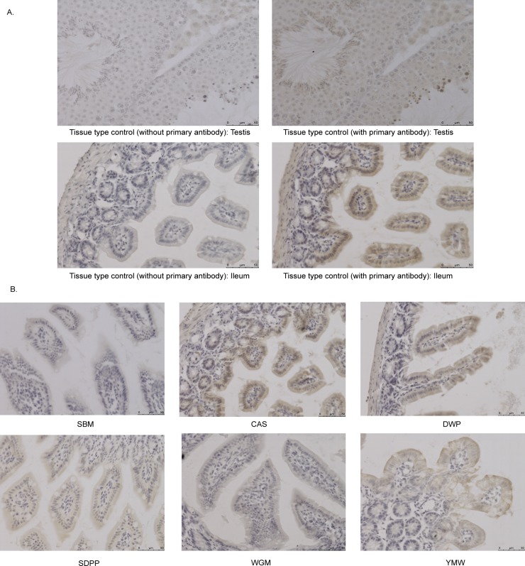
Fig 5. Immunohistochemistry of PFA-fixed paraffin-embedded tissue sections with anti-mTOR antibody in ileal tissue of mice fed with different experimental diets.
(A) Brown color indicates positive reaction in the image of tissue type control (testis) and a positive ileal tissue section (CAS). No positivity was observed in the same tissue by withholding the primary antibody (i.e. anti-mTOR antibody) during the staining procedure. (B) Immunohistochemistry of PFA-fixed paraffin-embedded ileum sections with anti-mTOR antibody of mice fed with different experimental diets. Brown color in the tissue section indicates positive reaction. SBM, soybean meal; CAS, casein; SDPP, spray dried porcine plasma; WGM, wheat gluten meal and YMW, yellow meal worm. Scale bar: 50 μm.

