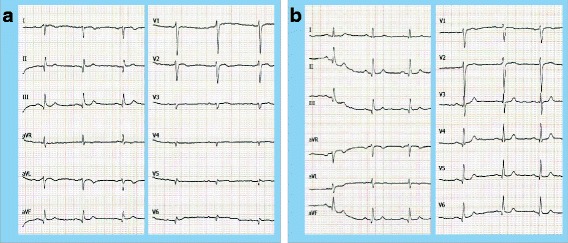
Fig. 1.

Electrocardiogram in dextrocardia (25 mm/s, 10 mm/mV). a Conventional placement of the ECG leads with the typical findings of dextrocardia: right axis deviation, positive QRS complexes (with upright P and T waves) in aVR, ‘global negativity’ (inverted P wave, negative QRS, inverted T wave) in I and absent R-wave progression in the chest wall leads. b Mirror inverted placement of the ECG leads on the right side of the chest and reversing the left and right arm leads
