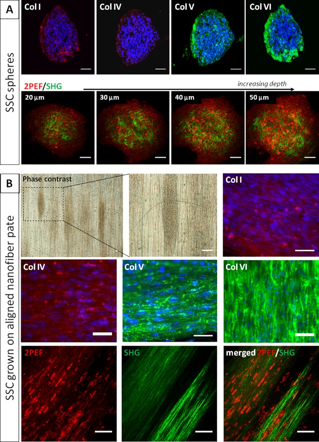
Fig 5. Collagen synthesis by cultured SSC.
(A) Spheres derived from SSC produce collagen type I, IV, V and VI (as shown by immunohistochemistry), with no apparent organization. As shown by multiphoton microscopy, these disorganized collagen fibrils (SHG signals in green) are present around cells (endogenous fluorescence, 2PEF in red) along the full depth of spheres of SSC. (B) When spheres of SSC were plated on nanofiber plates to promote synthesis of an organized collagen extracellular matrix, spheres tended to elongate along nanofibers (as shown by phase contrast microscopy). The same feature was evident after immunostaining of collagen type I, IV, V and VI. Aligned collagen fibrils (green) were also evidenced by SHG microscopy. Bars, 50 μm.

