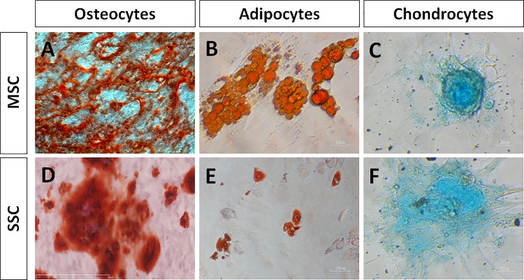
Fig 7. Mesenchymal cell differentiation assays of SSC.
After primary culture, MSC (used as positive controls) and SSC were dissociated and plated into osteocyte, adipocyte and chondrocyte differentiating media. MSC (A-C) and SSC (D-F) show the ability to differentiate into osteocytes identified by calcium deposits (A, D; Alizarin Red staining), adipocytes (B, F; lipids stained by Oil RedO) and chondrocytes (C, F; matrix proteoglycans revealed by Alcian Blue staining). Bars, 100μm.

