
Fig. 8.
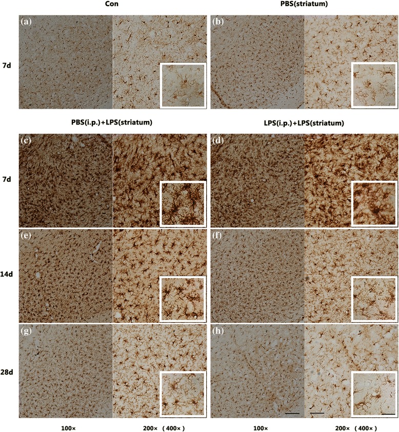
Comparison of microglial morphology in the SN among different pre-treated groups. The results displayed that intracranial LPS injection caused microglial activation respectively characterized by “amoeboid form” at 7 days (c, d) or “typical activated form” at 14 days (e, f) post to the LPS exposure. However, endotoxic tolerant preconditioning resulted in fewer microglial counting and lighter immunohistochemical staining (d, f, and h). Since the control group and the PBS (striatum) group were not statistically significant at each time point, we selected 7 days as a representative (a, b). Scale bars, 100 μm (100×), 50 μm (200×), and 25 μm (400×)