
Fig. 1.
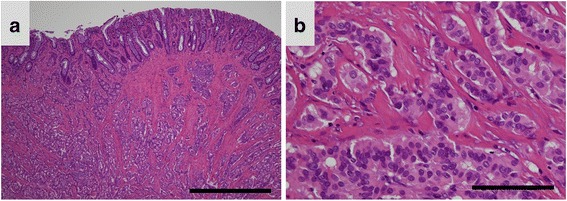
Representative images of histopathological findings in hindgut neuroendocrine tumors. a A photomicrograph showing a low-power field image of a hindgut neuroendocrine tumor (NET). The tumor cells are arranged in a trabecular pattern and show solid nests (Hematoxylin and eosin (HE) staining; original magnification, ×40; scale bar represents 1000 μm). b A photomicrograph showing a high-power field image of a hindgut NET. The tumor cells are uniform, arranged in rounded, solid nests, and have round-to-oval nuclei. Mild nuclear atypia can be seen (HE staining; original magnification, ×400; scale bar represents 100 μm)