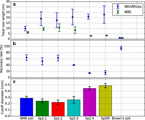
Fig. 4.

a Total root length extracted from Magnetic Resonance Imaging (MRI) data and after excavation and scanning in WinRhizo. b Total root length from MRI data relative to excavated root systems. For Sp2.4 and Sp5M, < 20% of the total root length was visualized in MRI. c Accordingly, the minimal root diameter detectable in MRI is significantly increased for Sp2.4 and Sp5M substrates compared to our lab standard NMR soil (Welch’s t test, p < 0.01). For Sp2.1, Sp2.2, and Sp2.3 we found no significant difference to NMR soil although a trend to lower cutoff values is visible. The limit could not be determined for Brown´s soil since here no thin roots were developed by the investigated plants. Error bars: ± 1 SD
