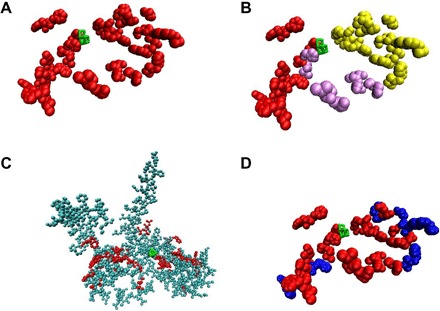
Fig. 3. Lumen-side oxidized residues detected in this study.

(A) Depiction of the 42 lumen-side oxidized residues detected in this study (red). (B) Same depiction as in (A), but arm 1 is shown in red, arm 2 in purple, and arm 3 in yellow. (C) All the 255 lumen-side residues that were covered by MS in this study, plus the covered and oxidized CP47 residues discussed in the text (see table S3). Some residues are obscured by others and are not visible in this view. Red, oxidized residues; cyan, nonoxidized residues. (D) Same depiction as in (A), except that in this view, the surface-exposed residues are colored blue, and buried residues are colored red. Visual Molecular Dynamics (VMD) (66) was used to determine whether a residue is surface-exposed or buried. Throughout the figure, the Mn cluster is shown in green.
