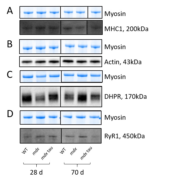
Representative blots for data shown in Table 2.

Shown for each panel is the myosin from the Stain Free gel, indicative of total protein (top) and the representative Western blot protein (bottom) for MHC1 (A, black line indicates non-contiguous lanes from the same gel), actin (B), DHPR (C) and RyR1 (D) in 28 d and 70 d WT, mdx and mdx tau mice.
