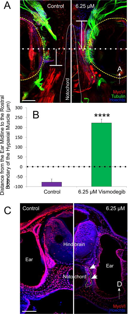
Figure 4. Rostral expansion of hypaxial muscle fibers following Vismodegib treatment.

A) The distance from the ear midline from the rostral boundary of the somites was calculated from animals immunostained with antibodies against tubulin (green), MyoVI (red) and counterstained with Hoechsts to label neurons, hair cells and muscle tissue, and nuclei, respectively. A line was drawn through the midline of the ear (dotted line), calculated from the anteroposterior diameter of the ear. From that line, the distance to the rostral boundary of the somites was determined (T-shaped line). Positive values were assigned for distances rostral to the midline and negative values for distances caudal to the midline. B) Means plus or minus standard errors of the mean for both controls and animals treated with 6.25 μM Vismodegib calculated using the method from (A). ****p<0.0001 C) Coronal section showing that hypaxial muscle fibers (outlined and arrows) expanded between the brain and the ear in animals treated with 6.25 μM Vismodegib. Scale bars represent 100μm.
