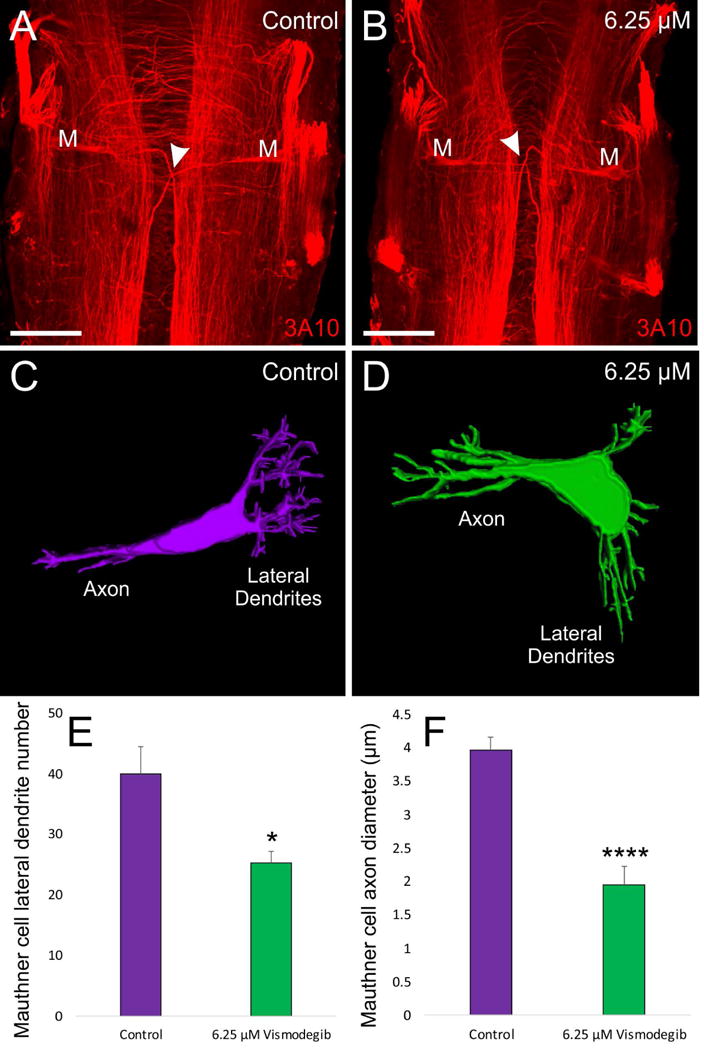
Figure 5. Effect of Vismodegib treatment on Mauthner Cell Development.

A) 3A10 antibody labeling of a pair of Mauthner cells from a control Xenopus.-B) 3A10 antibody labeling of a pair of Mauthner cells from an animal treated with 6.25 μM Vismodegib. Arrowheads for A and B indicate axon crossing at the midline. C) 3D reconstruction of a control Mauthner cell from dextran amine labeling D) 3D reconstruction of a Mauthner cell from an animal treated with 6.25 μM Vismodegib. E) Shh inhibition results in a significant reduction in the degree of branching of the Mauthner cell in 6.25 μM Vismodegib-treated animals (n = 4) compared to control animals (n = 4). F) Shh inhibition results in an approximately two-fold reduction in axonal diameter of the Mauthner cell in 6.25 μM Vismodegib-treated animals (n = 3) compared to control animals (n = 5). Scale bars represent 100 μm.* p<0.05, ****p<0.0001.
