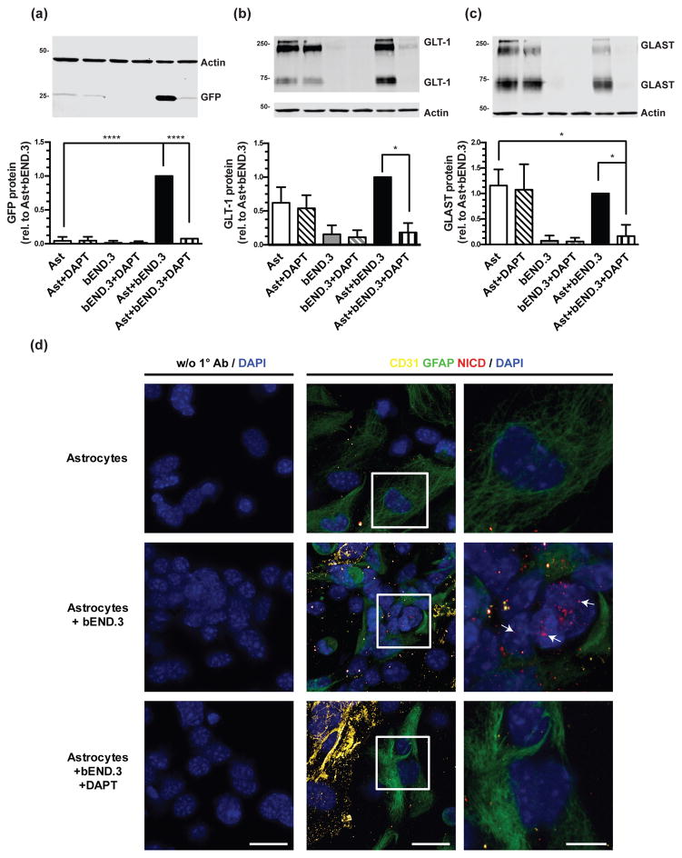
Figure 6. Effect of DAPT on eGFP, GLT-1, and GLAST protein levels and on Notch activation.
Cortical astrocytes from dual reporter mice were cultured alone or directly on top of an intact monolayer of bEND.3 cells and treated with 10μM DAPT for 10 days, and then harvested for Western blot analysis of eGFP (a), GLT-1 (b), and GLAST (c) protein levels. Top: Representative blots and Bottom: summary of quantification, normalized to β-actin. The β-actin blots are identical in panels (b) and (c), as GLT-1 and GLAST are quantified in the same immunoblot. Data are the mean ± SEM of three independent experiments. **** p ≤ 0.0001, * p ≤ 0.05 for indicated comparisons. (d) Expression of NICD was examined in cortical astrocytes from dual reporter mice, in astrocytes co-cultured with bEND.3 cells for 24 hours, and in co-cultures treated with 10μM DAPT. An anti-GFAP antibody was used as a marker of astrocytes, an anti-CD31 antibody was used as a marker of endothelia, and DAPI staining was used to identify nuclei. The magnification of the first two columns are the same (40x with 3x optical zoom, scale bar 25μm) and the last column is an 8x optical zoom of the region outlined by the white box in the middle column (scale bar 10μm). Representative of 3 independent experiments.

