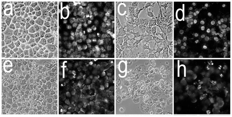
Figure 4.
Morphology and chromatin staining of A549 (a,b,e,f) and 1c1c7 (c,d,g,h) cells initially (a–d) and 16 h (e–h) after the combination PDT protocol. With the PDT conditions used, the clonogenic survival of A549 and 1c1c7 cells was ~20% and 10%, respectively. Phase contrast images (a,c,e,g) and Hö33342 fluorescence (b,d,f,h) are shown.

