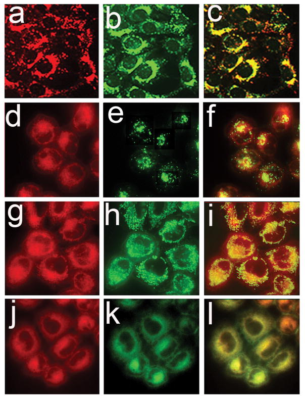
Figure 8.
Colocalization of NPe6 (top row) or BPD (bottom 3 rows) with fluorescent markers for lysosomes (top two rows), mitochondria (third row) and ER (bottom row) in A549 cell cultures. Cells were loaded with 20 μM NPe6 (a–c) or 0.5 μM BPD (d–l) for 1 h before imaging. Fluorescent probes for identification of lysosomes (LTG), mitochondria (MTG), and endoplasmic reticulum (ErTr) were added 15 min prior to imaging. Images for NPe6 (a) and BPD (d,g,j) fluorescence are shown along with fluorescence of LTG (b and e), MTG (h) and ErTr (k). Overlays of photosensitizer and probe fluorescence are shown in panels c,f,i and l.

