
Fig 2.
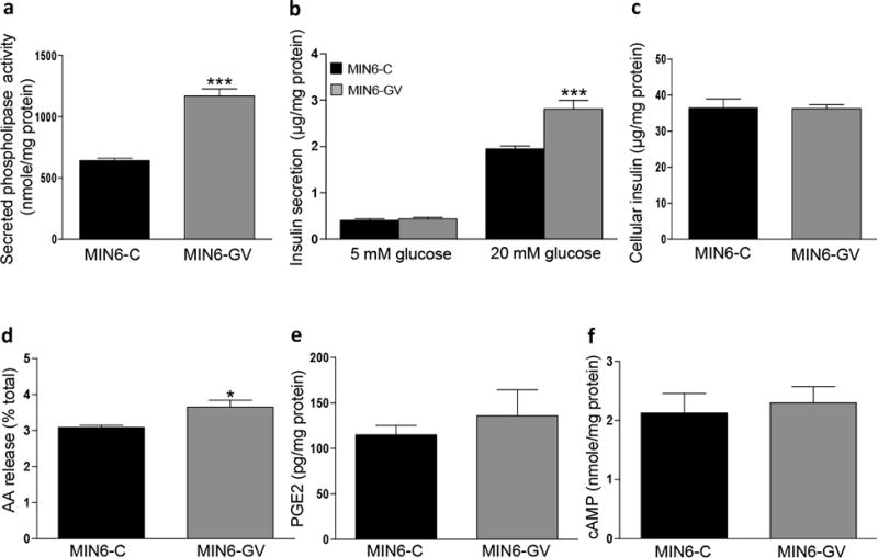
GV sPLA2 activates glucose-stimulated insulin secretion (GSIS) and enhances AA release in MIN6 cells. MIN6 cells were transfected with a control expression plasmid (MIN6-C) or an expression plasmid encoding GV sPLA2 (MIN6-GV) as described under “Materials and Methods.” a, Phospholipase activity in 48 h conditioned media from MIN6-C or MIN6-GV cell cultures was determined and normalized to cell protein. b, GSIS was performed in MIN6-C and MIN6-GV cells 48 h after transfection as described in “Materials and Methods.” Insulin levels in the media were determined and normalized to total cell protein. c, Total cellular insulin content of MIN6-C and MIN6-GV cells was assessed as described in “Materials and Methods.” d, [3H]-AA release by MIN6-C and MIN6-GV cells was quantified and expressed as the percent of total cellular [3H]-AA as described in “Materials and Methods.” e, PGE2 levels in culture media from MIN6-C and MIN6-GV cells 48 h after transfection, normalized to total cell protein. f, cellular cAMP content in MIN6-C and MIN6-GV cells was determined 48 h after transfection and normalized to total cell protein. Data are from 4 independent transfections per construct and are presented as mean ± S.E; *p<0.05. ***, p < 0.001.