Fig 5.

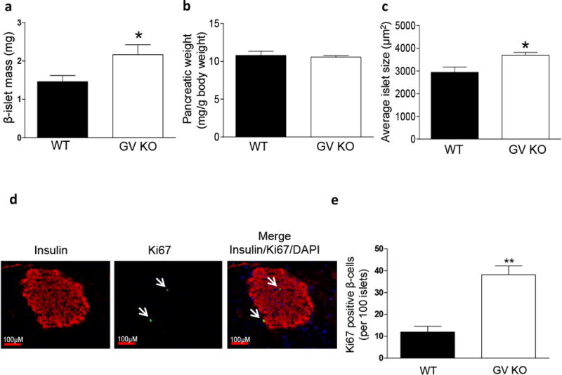
Increased β-islet mass, average islet size and β-cell proliferation in the pancreas of GV KO mice. a, Pancreatic β-islet mass of WT and GV KO mice (n=4/strain; 5-8 sections were analyzed from each mouse) were calculated by obtaining the fraction of the area of pancreatic tissue positive for insulin staining and multiplying this by the pancreatic weight as described in detail in the “Materials and Methods.” b, Pancreatic weight of WT and GV KO mice (n=4/strain). c, Average islet size of WT and GV KO pancreata (n=4/strain; 5-8 sections/mouse) was calculated by dividing the total area of pancreatic tissue positive for insulin staining by the total number of islets. d, Representative immunofluorescence images of pancreatic islets from GV KO mice at 20X magnification showing insulin (red) and Ki67 (indicated by arrows pointing to green staining). A merged image shows expression of Ki67 in insulin-producing cells of GV KO pancreas, co-localized with DAPI staining. e, Quantification of number of β-cells with Ki67-positive nuclei per 100 islets in WT and GV KO mice (n=3/group; 30-50 islets from 2 sections per mouse). Data are presented as mean ± S.E; *p<0.05; ***p<0.01.
