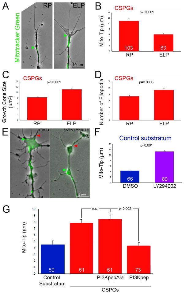
Figure 2.
Evidence for the involvement of the LAR receptor and suppression of PI3K signaling in mediating the effects of CSPGs on mitochondria positioning and growth cone morphology. (A) Examples of axons on CSPGs +/− treatment with the LAR inhibitory ELP peptide or the control inactive RP peptide(2 hrs). Phase contrast and Mitotracker green labeled mitochondria are shown. Labeling as in Figure 1B. (B) Graph showing Mito-Tip measurements under conditions of RP (control) and ELP treatment on CSPG substrata. Sample sizes showing the bars also apply to panels C and D. (C) Graph showing the effects of RP and ELP treatment on growth cone size on CSPG substrata. (D) Graph showing the effects of RP and ELP treatment on the number of growth cone filopodia on CSPG substrata. (E) Examples of growth cones on the control substratum treated for 30 min with DMSO or the PI3K inhibitor LY294002. Presentation as in panel (A). (F) Graph of Mito-Tip measurements for axons on the control substratum as a function of treatment with DMSO or LY294002. (G) Graph showing the effects of treatment with the PI3K activating PI3Kpep, and its inactive control PI3KpapAla, on Mito-Tip measurements. PI3KpepAla does not affect Mito-Tip measurements on CSPGs. However, treatment with PI3Kpep treatment decreases Mito-Tip measurements relative to RP treatment, and with PI3Kpep treatment Mito-Tip measurements are not different from the control substratum. Samples sizes are denoted in the bars.

