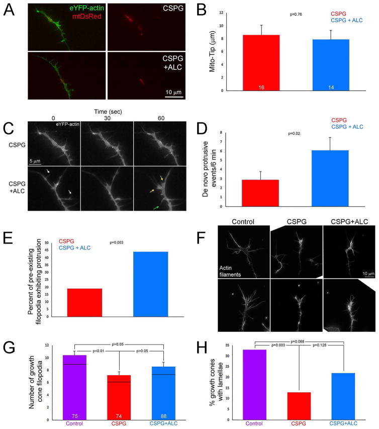
Figure 5.
Treatment with acetyl-L-carnitine (ALC) partially restores growth cone dynamics and morphology on CSPGs. (A) Examples of axons expressing eYFP-β-actin and mtDsRed on CSPG and CSPG+ALC. (B) Graph showing Mito-Tip measurements on CSPG and CSPG+ALC. (C) Examples of the dynamics of growth cones on CSPG + ALC. The two growth cones shown are the same as depicted in panel A. The growth cone on CSPGs exhibits very little dynamics as reflected by minimal remodeling. In contrast, the growth cone treated with ALC is much more dynamic. Examples of lamellar protrusion (30–60 sec, yellow arrows at 60 sec) and filopodial protrusion (30–60 sec, green arrow at 60 sec) are shown. Also note that not only was protrusive activity increased at growth cones but also retraction of filopodia and lamellae (white arrows at 0 sec). (D) Graph showing de novo protrusive events at growth cones in the form of filopodia or lamellipodia on CSPG and CSPG+ALC. (E) Graph showing the percentage of filopodia existing at the beginning of the video sequence that undergo > 1 μm protrusion on CSPG and CSPG+ALC. (F) Examples of growth cones stained with rhodamine phalloidin to reveal actin filaments on the control substratum, CSPG and CSPG+ALC. (G) Graph showing the number of filopodia at growth cones on the control substratum, CSPG and CSPG+ALC. (H) Graph of the percentage of growth cones exhibiting one or more lamellae on the control substratum, CSPG and CSPG+ALC. Bars are color coded as in panel G. Samples sizes are denoted in the bars.

