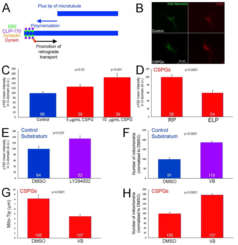
Figure 7.
Elevated levels of p150-Glued in distal axons on CSPG substrata. (A) Schematic of the mechanism that drives the initiation of retrograde transport away from distal axons. Briefly, microtubule plus tips undergoing polymerization recruit EB3 that in turn serves to scaffold a complex consisting of CLIP-170, dynactin and dynein. This complex then promotes the activation of retrograde organelle evacuation from growth cones. Thus, impairing microtubule plus tip dynamics, or the function of dynein-dynactin, prevents to initiation of retrograde transport from the distal axon. (B) Examples of p150glu dynactin staining in growth cones on the control and CSPG (10 μg/mL) substrata. (C) Quantification of the staining intensity of p150Glu staining in growth cones as a function of CSPG concentrations used to coat substrata. Comparison to the control substratum. (D) Culturing in the presence of ELP decreases the staining intensity of p150Glu in growth cones on 5 μg/mL CSPGs relative to the control treatment with RP. (E) Inhibition of PI3K activity using LY294002 (25 μM) increases the staining intensity of p150Glu in growth cones on the control substratum. (F) Suppression of microtubule plus tip dynamics using 3 nM vinblastine (VB; 60 min) increases the number of mitochondria in the distal 10 μm of axons on control substrata. (G) Treatment with vinblastine (60 min) on CSPG substrata decreased Mito-Tip. (H) Treatment with vinblastine on CSPG substrata increased the number of mitochondria in distal axons. Samples sizes are denoted in the bars.

