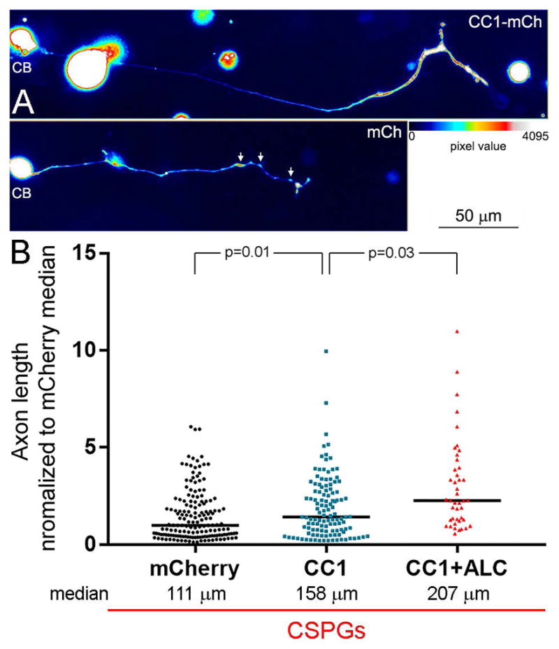
Figure 8.

Disruption of the interaction between dynactin and dynein increases axon lengths on CSPG substrata. (A) False colored examples of neurons (CB = cell body) and their axons on CSPGs expressing CC1-mCherry (mCh) or mCh alone. The distal axon of the CC1-mCh expressing neurons exhibits distal thickening as represented by warmer colors in the false colored image (see color bar). The arrows in the mCh panel show the presence of varicosities along the distal axon, as previously detailed in Figure 1, also evidenced by warmer colors. (B) Graph of axon length measurements for mCh, CC1-mCh and CC1-mCh plus 500 μM acetyl-L-carnitine (ALC). Black lines in bars denote the medians as on CSPGs axon length measurements were not normally distributed. Samples sizes are denoted in the bars.
