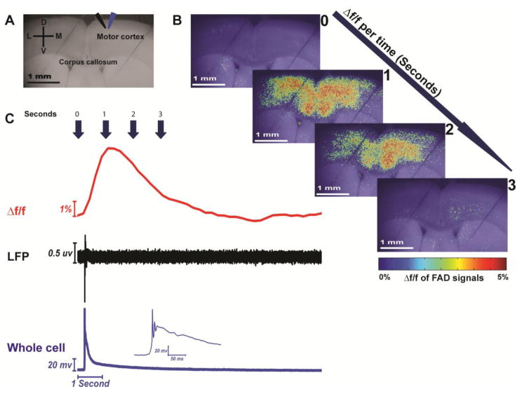
Figure 3. Simultaneous flavoprotein imaging and electrophysiological recording of the spontaneous paroxysmal depolarizing (SPD) events evoked by SR-95531.
A) Grayscale image of coronal brain slice showing the site of LFP (black arrow) and whole-cell recording (blue arrow); B) Pseudocolor heat maps of coronal brain slice showing the change of Δf/f of FAD signals associated with SPD events per time; C) Time traces of Δf/f of FAD (top panel), LFP (middle panel), and whole cell recording (bottom panel) signals of SPD events occurring in the presence of 4 μM SR-95531.

