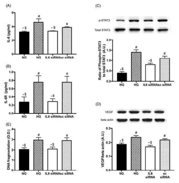
Figure 5.
The role of IL-6 signaling on STAT3/VEGF pathway and apoptosis. REC were cultured in normal glucose (5 mM, NG) or high glucose (25 mM, HG) and transfected with human IL-6 siRNA (20 nM of final concentration). ELISA results showed that the levels of both IL-6 (A) and sIL-6R (B) in REC were suppressed by IL-6 siRNA in high glucose conditions. (C and D) STAT3 phosphorylation and VEGF levels were significantly reduced by inhibited IL-6 signaling. A representative blot is shown. (E) ELISA data showing decreased levels of DNA fragmentation in REC. #p < 0.05 versus NG, *p < 0.05 versus HG, $p < 0.05 versus Neg.; N=4 (A, B, and D), N=7 (C and E); Data are mean±S.E.M.

