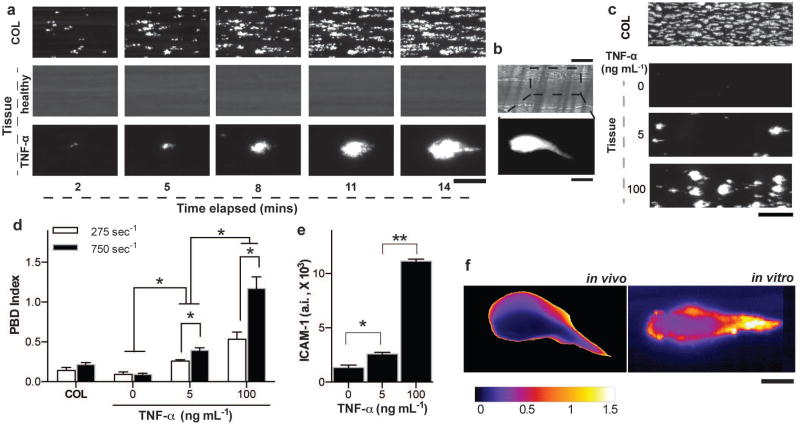
Figure 2. Analysis of dynamical progression of thrombus formation in the alveolus chip.
a) Fluorescence micrographs depicting a section of the imaged microchannel (Scale bar, 25 µm) showing platelet accumulation over time (left to right) on collagen (top), a healthy blood vessel (middle), TNF-α stimulated vessel (bottom) and b) platelet accumulation after 4 minutes of laser-induced injury on a mouse cremaster arteriole (top, bright field and bottom, fluorescence; Scale bar, 25 µm, top and bottom). c) Fluorescent micrographs of a large section of the vascular chamber showing luminal thrombus formation on bare collagen (topmost) and TNF-α stimulated endothelium in a dose dependent manner (bottom three; Scale bar, 100 µm). d) Sensitivity analysis of the platelet-endothelial dynamics algorithm (PBD index), showing that in conditions of vascular injury/collagen or healthy endothelium, the dynamical events characterizing clot formation are nearly absent but they increase in a TNF-α dose dependent manner. The PBD index is also sensitive to applied shear rate (n = 3, *P<0.05, 2-way ANOVA). e) ICAM-1 expression on the endothelial cells after stimulation of the vessel with TNF-α (n = 4, *P<0.05, unpaired t-test). f) Image colormap showing the coefficient of variability (calculated from the fluorescence image timeseries) within a laser-induced thrombus in vivo (left) and TNF-α induced thrombus on a vsacular lumen in vitro (right). Scale bar, 20 µm.

