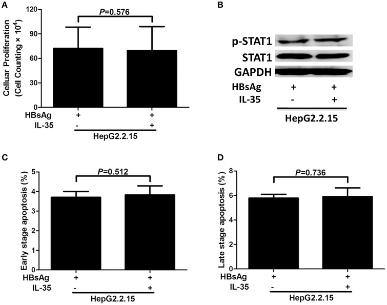
Figure 3.
The regulatory role of interleukin (IL)-35 in HepG2.2.15 cells. HepG2.2.15 cells were stimulated recombinant hepatitis B surface antigen (HBsAg) in the presence or absence of recombinant IL-35 for 24 h, and were performed independently for five times. (A) Cellular proliferation was measured by cell counting kit-8. The data were presented as mean ± SD, and significances were calculated using paired t-test. (B) Phosphorylated STAT1 (p-STAT1) and total STAT1 were tested by Western blot, and GADPH was shown as control. The percentages of early stage apoptotic cells (C) and late stage apoptotic cells (D) were shown. The data were presented as mean ± SD, and significances were calculated using paired t-test.

