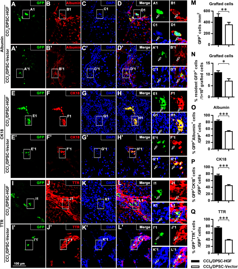
Figure 4.
HGF overexpression promotes the hepatocyte-like differentiation of grafted DPSCs. (A–L,A’–L’) Fluorescence microscopy images of hepatic tissue immunostained with anti-GFP (A,A’,E,E’,I,I’), anti-albumin (B,B’), anti-CK18 (F,F’), and anti-TTR (J,J’) and counterstained with DAPI (C,C’,G,G’,K,K’) and related merged fluorescence microscopy images (D,D’,H,H’,L,L’) in CCl4/DPSC-HGF (A–L) and CCl4/DPSC-HGF groups (A’–L’). A1-L1 and A’1-L’1 show magnified images of the outlined areas from the corresponding panels. (M-Q) Quantitative analyses of the number of GFP+ graft-derived cells (M), the percentage of resident GFP+ cells in total 1 × 10⁶ grafted cells (N) and the percentages of albumin+ (O), CK18+ (P) and TTR+ (Q) cells in the GFP+ graft-derived cell population. Scale bar (A–L,A’–L’) = 100 μm. **P < 0.01; ***P < 0.001.

