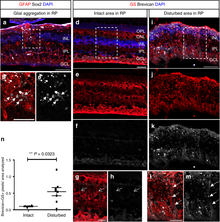
Fig. 7.
Brevican in MG of disorganized human retinitis pigmentosa retinas. a–m Immunofluorescence for Tomato, GFAP, and Sox2 (a–c) or glutamine synthetase (GS) and Brevican (d–m), as well as DAPI nuclear staining of retinal cross sections of retinitis pigmentosa patients. Glial aggregations (white arrows in b, c) found in the INL resemble to those found in the 6-month-old Dicer-CKOMG retinas. MG in intact regions (arrows in g, h) were Brevican– while glial aggregations found in disturbed regions (arrows in l, m) were Brevican+. n Brevican/GS immunoreactivity assessed in intact (n = 4) and disturbed (n = 8) retinitis pigmentosa samples showed a significant increase in Brevican protein level in the disturbed samples. Statistics: mean ± S.E.M., independent samples t-test and Levene’s test for equality of variances, 2-tailed, p = 0.0323. Scale bars in a, i: 100 μm, in b, d: 50 μm in g, l: 25 μm. ONL, OPL, INL, IPL, GCL, NFL as defined in Fig. 1 legend, RP retinitis pigmentosa

