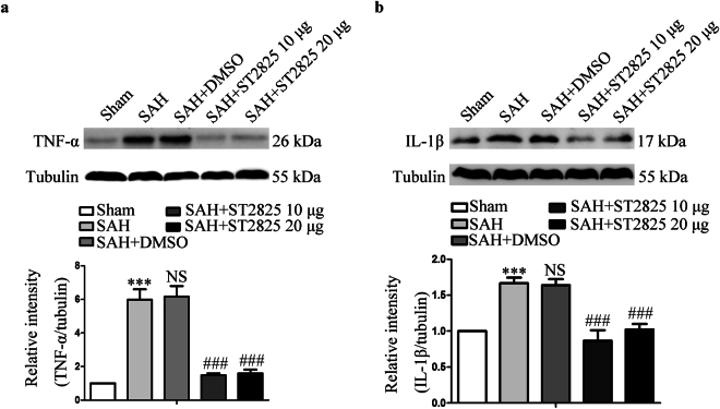
Figure 5.
Effects of MyD88 inhibition on TNF-α and IL-1β levels 24 h post-SAH. A substantial increase in TNF-α (a) and IL-1β (b) was found in anterior basal temporal lobes from SAH rats 24 h after SAH. MyD88 inhibition successfully reduced SAH-induced increase of TNF-α and IL-1β expression. The upper panel shows representative protein levels of TNF-α and IL-1β. The bottom panels are quantitative data. Tubulin was used as loading control. Data are expressed as mean ± SD (n = 6 in each group). TNF-α, tumor necrosis factor-α, IL-1β, Interleukin-1β, Definition of SAH is the same as in Fig. 2. ***p < 0.001 compared with the sham group, ###p < 0.001 compared with the SAH group, NS, no statistic difference compared with the SAH group.

