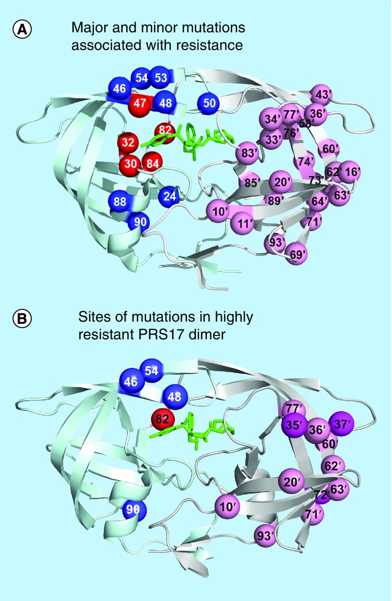
Figure 2. . Protease dimer showing sites of resistance mutations.
(A) Major and minor mutations associated with resistance. Locations of resistance-associated mutations mapped on the HIV protease dimer bound with DRV (PDB: 2IEN). The protease dimer is shown in ribbons with DRV in black sticks. Sites of mutations associated with drug resistance [20] are indicated by spheres. Mutations altering inhibitor binding are in dark gray and mutations affecting dimer stability or flap dynamics are indicated in light gray on the left subunit. Distal mutations with poorly defined effects are shown in gray on the right subunit.
DRV: Darunavir.

