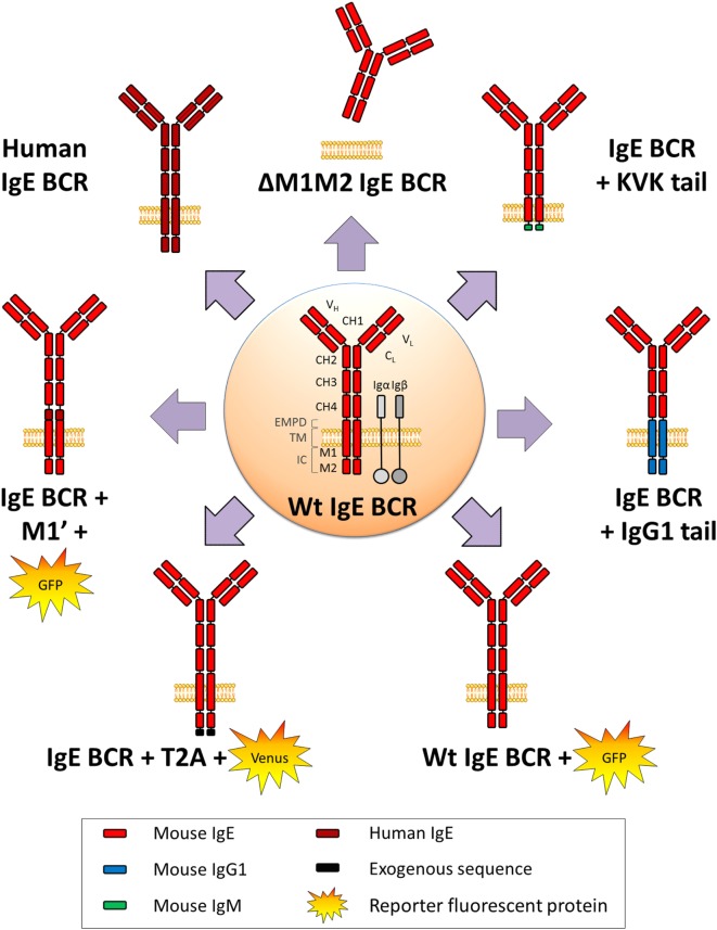
Figure 3.
WT and variant IgE B cell receptor (BCR) expressed in mouse models. Mouse WT IgE BCR structure is schematized in the center. Heavy and light chain variable (VH and VL) domains form the antigen-binding site, constant domains (CH), transmembrane (TM), and intracellular domains (IC) are shown, and anchored in the lipid membrane. IgE BCR includes an extra-membrane proximal domain (EMPD). Co-signaling molecules Igα and Igβ are represented in gray. Variant and chimeric IgE BCR resulting from the different knock-in and knock-out alleles (described in Figure 2) are represented, and reporter fluorescent proteins are illustrated.

