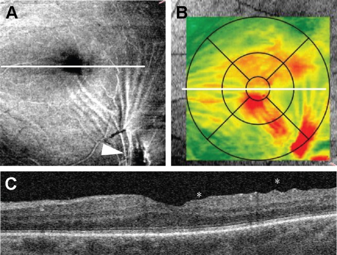
Figure 2.

Retinal wrinkling associated with eccentric epiretinal membrane. (A) En face image demonstrates epiretinal membrane (arrowhead) and tension lines spreading along the inner retinal surface to the foveal center. (B) Retinal thickness map shows retinal thickening within central subfield. (C) B-scan through the foveal center demonstrates retinal wrinkling (asterisks). The white line represents the position of B-scan.
