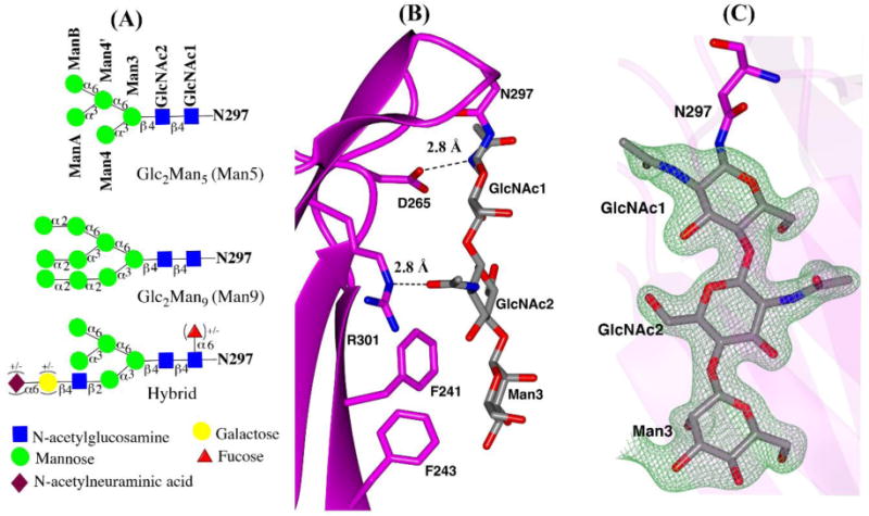
Fig. 4.

N-glycans in IgG3 Fc structure (A) schematic representation of Man5GlcNAc2 (Man5), Man9GlcNAc2 (Man9) and hybrid glycoform based on the Essentials system of glycan nomenclature (B) close-up view of N297 attached glycans and protein contacts. H-bonds between protein and carbohydrate are shown as black dotted lines. (C) ordered glycans from chain A in stick representation, shown with Fo-Fc electron density difference map contoured at 3σ.
