
Figure 1. Lkb1 is expressed in the granule cell precursors.
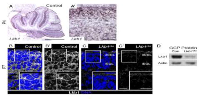
A–A′. Lkb1 in situ hybridization at postnatal day 4 (P4). Lkb1 is expressed in all cortical layers but is highest in the external granule layer (EGL). B–C. Immunohistochemistry for Lkb1 (white) and TO-PRO 3 (blue) at postnatal day 7 (P7). Lkb1 localizes to the cytoplasm and cell cortex of GCPs in the EGL of control cerebella (B–B′) but is absent in Lkb1cko cerebella (C–C′). D. Western blotting for Lkb1 reveals a significant reduction in Lkb1 protein levels in Lkb1cko GCPs compared to controls. Actin was used as loading control. Scalebars: A = 500 μm, A′ = 50 μm, C–D = 10 μm. Con = control, EGL = external granule layer, oEGL = outer EGL, iEGL = inner EGL, ML = molecular layer, IGL = internal granule layer. Full-length blots are shown in Supplementary Figure 9.