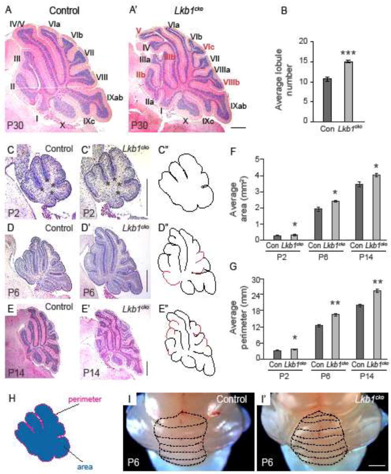
Figure 2. Granule cell precursor-specific loss of Lkb1 results in increased foliation and cortical expansion.

A–A′. Hematoxylin and eosin staining of control (A) and Lkb1cko (A′) cerebella at P30. Roman numerals denote lobule numbers. Red roman numerals indicate lobules present in Lkb1cko absent in control. B. Average lobule number of control and Lkb1cko cerebella. C–E. Hematoxylin and eosin staining of mid-vermal cerebellar cross sections at the indicated stages. Lobules present in Lkb1cko not present in the control are highlighted in red in C″–E″. Asterisks in C–C′ indicate cardinal fissures. F. Average cross sectional area of mid-vermal cerebellar sections at indicated stages. G. Average cross sectional perimeter of mid-vermal cerebellar cross sections at the indicated stages. H. Illustration showing how area and perimeter were determined. I–I′. Whole mount images of P6 control (I) and Lkb1cko (I′) cerebella. Dashed lines delineate folia. n=5 for all analyses. *, p<0.05, **, p<0.005, ***, p<0.0005, Student’s t-test. Scalebar 500 μm for all images. Con = control.
