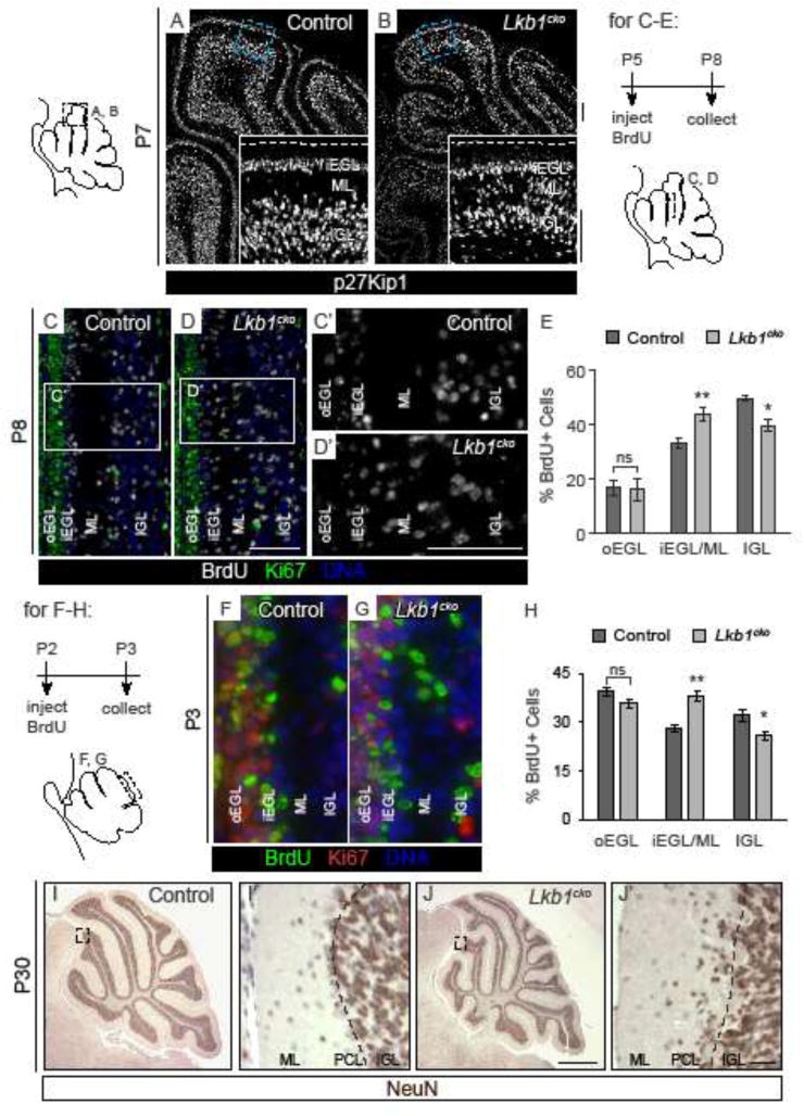
Figure 6. Lkb1cko cerebella have defects in granule cell migration.

A–B. p27Kip1 immunostaining labels post-mitotic GCPs in P7 control (A) and Lkb1cko (B) cerebella. Dashed line in inset denotes cerebellar surface. C–D. BrdU/Ki67 co-staining of P8 control (C) and Lkb1cko (D) cerebella three days after BrdU injection. C′–D′. Enlarged images of boxed regions in C and D. E. Quantification of the proportion of BrdU+ cells in each of the specified regions three days after BrdU pulse. n=3 controls, n=5 Lkb1cko. *, p<0.05, ** p<0.005. Student’s t-test. F–G. P3 cerebella injected with BrdU at P2. H. Quantification of the proportion of BrdU+ cells in each of the specified regions one day after BrdU pulse. n=3, *, p<0.05, ** p<0.01. Student’s t-test. I–J. Representative staining for Neuron-specific nuclear protein (NeuN), a marker of mature granule cells, in P30 control (I) and Lkb1cko (J) cerebella. I′ and J′ are enlargements of the boxed regions in I and J. Dashed lines in I′ and J′ corresponds to Purkinje cell layer (PCL). Note that a number of granule cells fail to migrate past the Purkinje cell layer in Lkb1cko. All scalebars 50 μm except I–J, in which scalebar is 500 μm. oEGL = outer external granule layer, iEGL = inner external granule layer, ML = molecular layer, IGL = internal granule layer. Full-length blots are shown in Supplementary Figure 9.
