
Fig. 2.
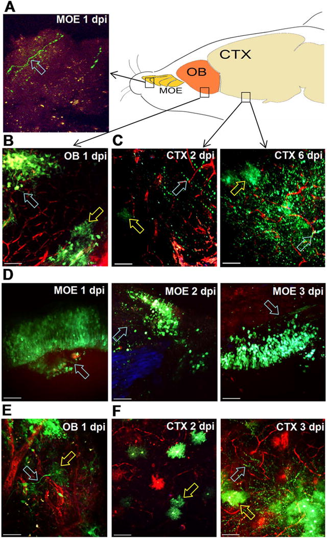
Intranasal VEEV neural invasion and dissemination in wild-type versus Ifit1−/− mice. A–C. Wild-type and Ifit1−/− mice were challenged with 107 PFU.n. VEEV TC83-GFP and tissues explanted for 2P microscopy at the indicated times. 70 kD TMR-dextran was injected i.v. to visualize the brain vasculature (red). In wild-type animals, infected cells (green) were detected in (A) the main olfactory epithelium (MOE) with olfactory neuron morphology (cyan arrow) 1 dpi and (B) olfactory bulb (OB) clustering in glomerular structures (cyan arrow) and nearby cells with astrocyte morphology (yellow arrow) on 1 dpi. C. Infected cells (green) in the cortex (CTX) 2 dpi have both neuronal (cyan arrow) and microglial or astrocyte morphology (yellow arrow). At 6 dpi, TC83-GFP reporter virus is widespread in the cortex and infected cells are observed with both neuronal (cyan arrow) and microglial morphology (yellow arrow) D. In Ifit1−/− mice, infected cells (green) have olfactory sensory neuron morphology (cyan arrows) and are wide spread in the MOE over the first three days post infection. Infected neuronal tracks projecting towards the cribriform plate were also observed (right panel, cyan arrow). TC83-GFP cells with different neuronal and astrocyte/microglia (cyan, yellow arrows) were widespread in Ifit1−/− mice (E) in the OB at 1 dpi and (F) the cortex at 2 and 3 dpi. Scale bar = 50 μm. (For interpretation of the references to color in this figure legend, the reader is referred to the web version of this article.)