Figure 4.
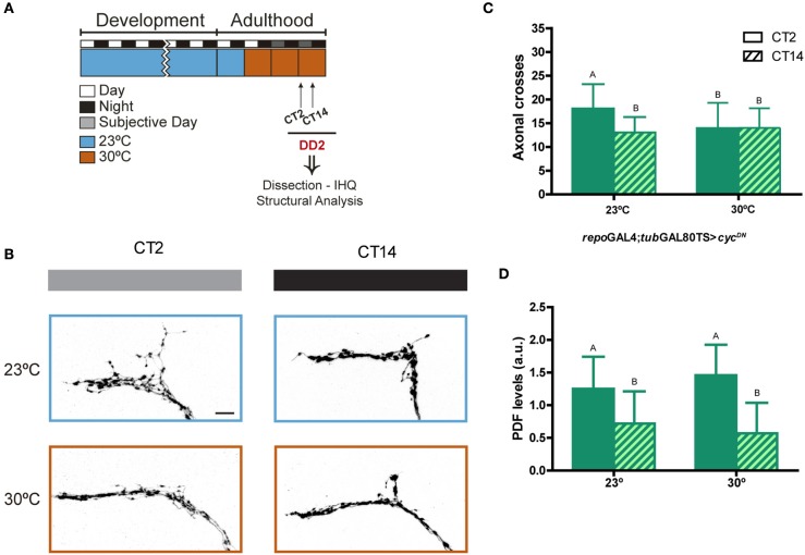
Clocks in glia are required for circadian remodeling of neuronal terminals. (A) Schematic diagram illustrating the standard protocol; the restrictive condition is highlighted in light-blue (23°C), and the permissive condition is shown in orange (30°C). (B) Representative confocal images of dsRed immunoreactivity at the dorsal protocerebrum of flies containing UAS-cycDN under repo-Gal4;tub-Gal80TS; pdf RED enables visualization of the axonal terminals. Brains were dissected at the early subjective day (CT2, gray bar) and early subjective night (CT14, black bar) during the 2nd day of constant darkness (DD2), which corresponds to the 3rd day of permissive condition (30°C). Control flies (always maintained at 23°C) are indicated in light-blue. (C) Quantitation of total axonal crosses of repo-Gal4;tub-Gal80TS > cycDN. Control flies (kept at 23°C) display circadian structural remodeling of axonal terminals while animals induced at 30°C show no differences along the day. Data represents the average of 3 experiments; a minimum of 27 brains were analyzed per CT/genotype. Different letters indicate statistical differences with a p < 0.05 (Two-way ANOVA with a Tukey post-hoc test, n = 8–10, N = 3). (D) Quantitation of PDF immunoreactivity at the dorsal protocerebrum from brains dissected at CT2 and CT14 on DD3. Control flies (23°C), exhibit circadian oscillation of PDF levels; those expressing cycDN at 30°C were not significantly different from controls. Same letters indicate no statistically different conditions (p > 0.05) (Kruskal–Wallis One-way ANOVA, followed by a Conover post-hoc test, n = 8–10, N = 2). Data represents the average of 2 experiments; a minimum of 16 brains were analyzed per CT/genotype.

