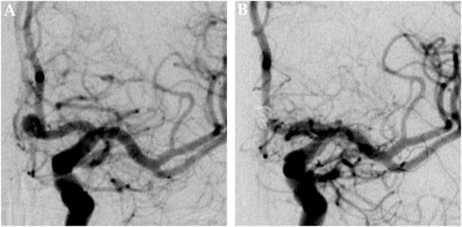
Figure 1.
Saccular aneurysm of the left anterior cerebral artery. Panel (A) shows a pretreatment DSA image of a saccular aneurysm at the A1–A2 junction of the left ACA in posterior–anterior projection. Panel (B) shows the corresponding 6 months posttreatment control DSA image, the aneurysm is completely excluded from the intracranial circulation; the formerly aneurysm-carrying vessel displays regular endoluminal contrast filling and now reveals signs of periprocedural damage.

