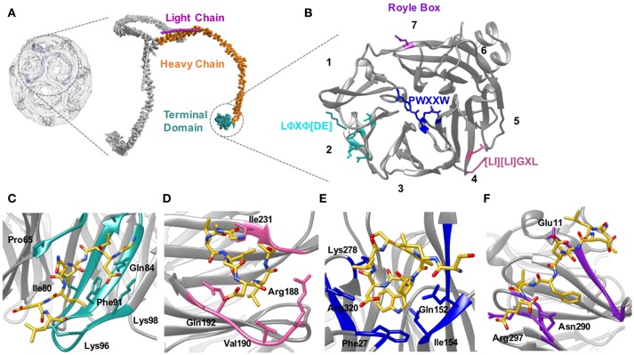
Figure 5.
Location of the clathrin heavy chain N-terminal domain (TD) and the location of the adaptor binding sites. (A) Clathrin forms a polymerized lattice structure around the growing vesicle in concert with adaptor proteins. The functional monomer, the triskelion, is formed of a trimer of heavy chains (~190 kDa, orange) with a smaller light chain (~25 kDa Pink) located along the top edge near the dimerization domain. The TD (cyan) is the primary binding location for adaptor proteins, and is located on the inside of the cage closest to the plasma membrane. (B) The TD is a 7-bladed β propellar that has 4 known sites for binding adaptor proteins. Site 1 between blades 1 and 2 is known as the clathrin box site (LΦXΦ[DE]), (turquoise); Site 2 situated in the center of the propellar is known as the W-box (PWXXW), (blue); Site 3 is the Arrestin-box ([LI][LI]GXL), (Pink); and Site 4 is the Royle Box which as yet has no defined interaction sequence, (purple). The numbers indicate the blade number. Peptides or protein bound with the 4 sites are indicated in the following four panels: (C) Clathrin box site of β2 adaptin (CGDLLNLDLG) bound to site 1; (D) βarrestin1L peptide (ALDGLLGG) bound to site 3; (E) amphiphysin peptide (TLPWDLWTT) bound to site 2; (F) an amphiphysin peptide bound to site 4. Structures are derived from PDB codes: 3IYV (A) (Fotin et al., 2004), 5M5R (B,C) (Muenzner et al., 2017), 3GD1 (D) (Kang et al., 2009), 1UTC (E) (Miele et al., 2004), 5M5T (F) (Muenzner et al., 2017).

