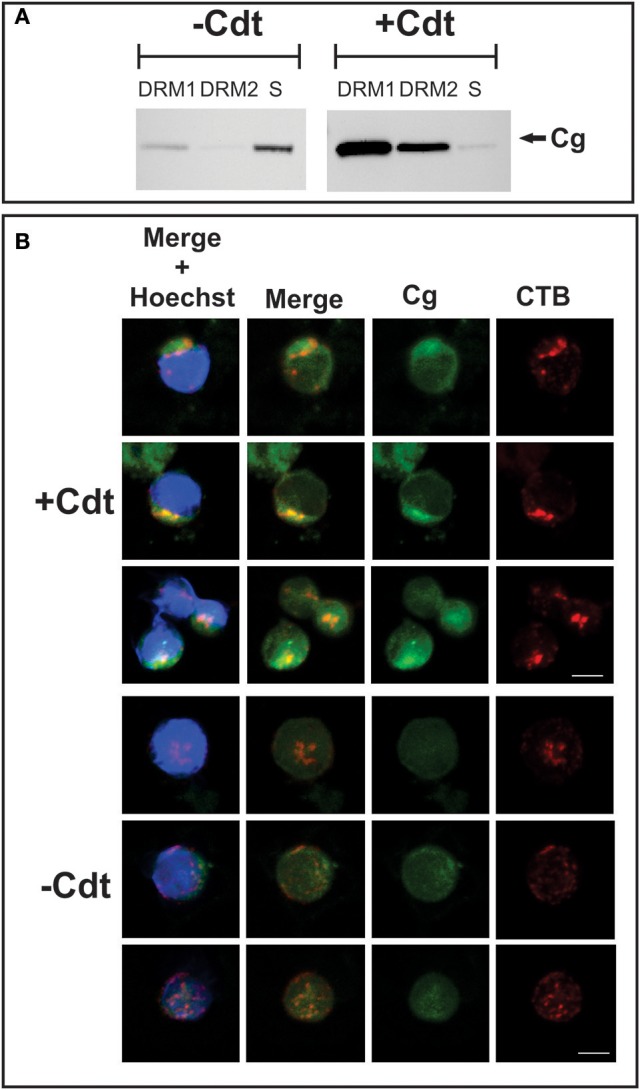
Figure 2.

Translocation of cellugyrin to cholesterol rich micro domains. (A) Jurkat cells were treated with medium (−Cdt) or with 2 μg/ml Cdt for 2 h. Cells were harvested, washed, and cholesterol rich microdomains isolated as detergent resistant membranes (DRM) as described in Materials and Methods. Two DRM zones, designated DRM1 and DRM2, as well as a soluble fraction were obtained and further analyzed by Western blot for the presence of cellugyrin. Results are representative of three experiments. (B) Jurkat cells were treated with medium (−Cdt) or 1 μg/ml Cdt (+Cdt) for 1 h; cells were stained and fixed as described in Materials and Methods and analyzed by confocal microscopy. Maximum intensity projection of a 3 μm z-stack series is presented (3 cells/condition). For each image, fluorescence is shown for cellugyrin alone (green), lipid rafts using fluorescence of cholera toxin B (CTB; red) and merged images (yellow) with (blue) and without nuclear staining Results are representative of multiple fields and analysis of over 50 cells for each condition. Scale bar = 5 μm.
