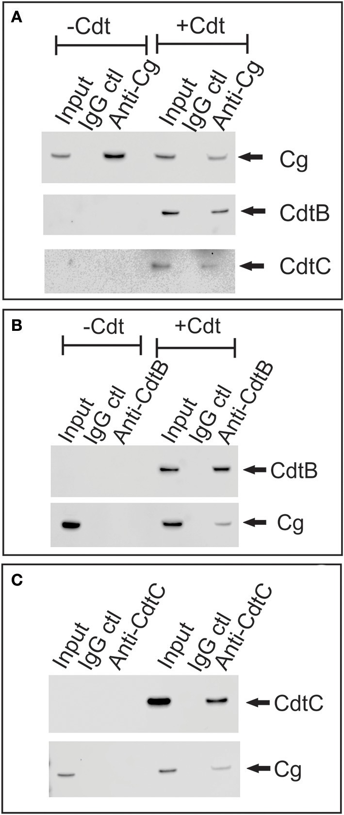
Figure 3.

Immunoprecipitation of cellugyrin and Cdt subunits. Jurkat cells were treated with medium or Cdt (2 μg/ml) for 2 h and then washed and homogenized as described in Materials and Methods. (A) Shows the results of extracts immunoprecipitated with either immobilized control IgG or anti-cellugyrin antibody. The bound material was eluted and analyzed by Western blot for the presence of cellugyrin (Cg), CdtB or CdtC. (B) Shows the results of cell extracts obtained from similarly treated cells as above and immunoprecipitated with immobilized control IgG or anti-CdtB mAb. The bound material was eluted and analyzed by Western blot for the presence of CdtB and Cg. (C) Shows the results of cell extracts obtained from cells treated as described above and immunopreciptated with immobilized control IgG or anti-CdtC mAb. The bound material was eluted and analyzed by Western blot for the presence of CdtC and Cg. Results are representative of three experiments.
