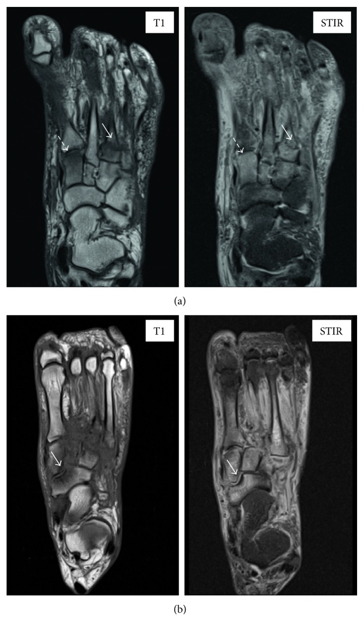
Figure 2.

Noncontrast large field-of-view MR scans in active Charcot osteoarthropathy. Representative examples of bone marrow oedema at the medial cuneiform (BMO score = 2; white dashed arrow) and fracture at the base of the 3rd metatarsal (fracture score = 1; white arrow) noted on axial T1 and STIR MR images (a). Example of collapse of the navicular bone (fracture score = 2, white arrow) noted on axial T1 and STIR MR images (b).
