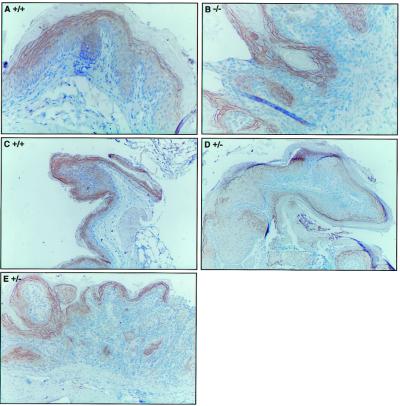
Figure 3.
Immunohistochemical detection of Fhit in NMBA-treated forestomach tissue. Forestomach sections were analyzed for expression of Fhit protein by immunohistochemistry using the rabbit anti- glutathione S-transferase-Fhit antiserum. (A) Normal epithelium in +/+ mouse 53; (B) basal cell hyperplasia in −/− mouse 16; (C) early papilloma in +/+ mouse 53; (D) early papilloma in +/− mouse 55; and (E) squamous cell carcinoma in +/− mouse 62. Note that the staining procedure results in nonspecific staining (B) of the highly keratinized outer layer. (Magnifications: A and B, ×200; C–E, ×100.)

