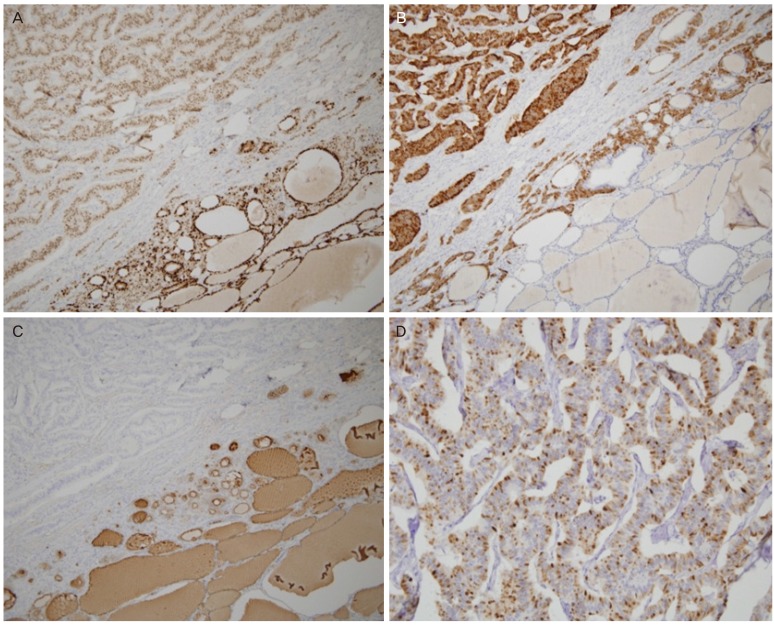
Fig. 2.
Immunohistochemical staining. (A) The tumor cells are diffusely positive for PAX8. (B) Carcinoid part of the tumor shows positive immunoreactivity for synaptophysin. (C) Follicles are surrounded by cells immunohistochemically positive for thyroglobulin. (D) Most of the cytoplasm of carcinoid tumor cells is positive for peptide YY.

