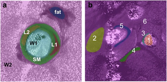
Fig. 6.

(a) Six anatomical compartments segmented for SLAM reconstruction overlaid on a T1-weighted IV image from a diseased vessel specimen (fat; two lesions, L1 and L2; vessel fluid contents, W1; smooth vessel-wall muscle, SM; surrounding tissue = W2). (b) In vivo SLAM segmentation of six compartments on an in vivo T1 image of the inferior vena cava (1, blood; 2, surrounding tissue; 3, arterial wall; 4, fat stripe; 5, vein wall; 6, everything else)
