
Figure 1.
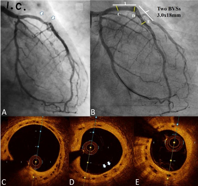
A: Coronary angiography (CAG) of the left coronary artery (LCA): Proximal to mid segment of left anterior descending artery (LAD) showed diffuse diseased lesions (white arrows); B: CAG of LCA: 3.5-year follow-up CAG showed fair coronary flow without stenosis; (C, D, and E) Optical coherence tomography (OCT) of LAD showed complete neointimal coverage of the residual bioresorbable vascular scaffold (BVS) struts (black boxes) from proximal to mid LAD (C-E). D: The overlap site showed two layers of BVS struts (white arrows).